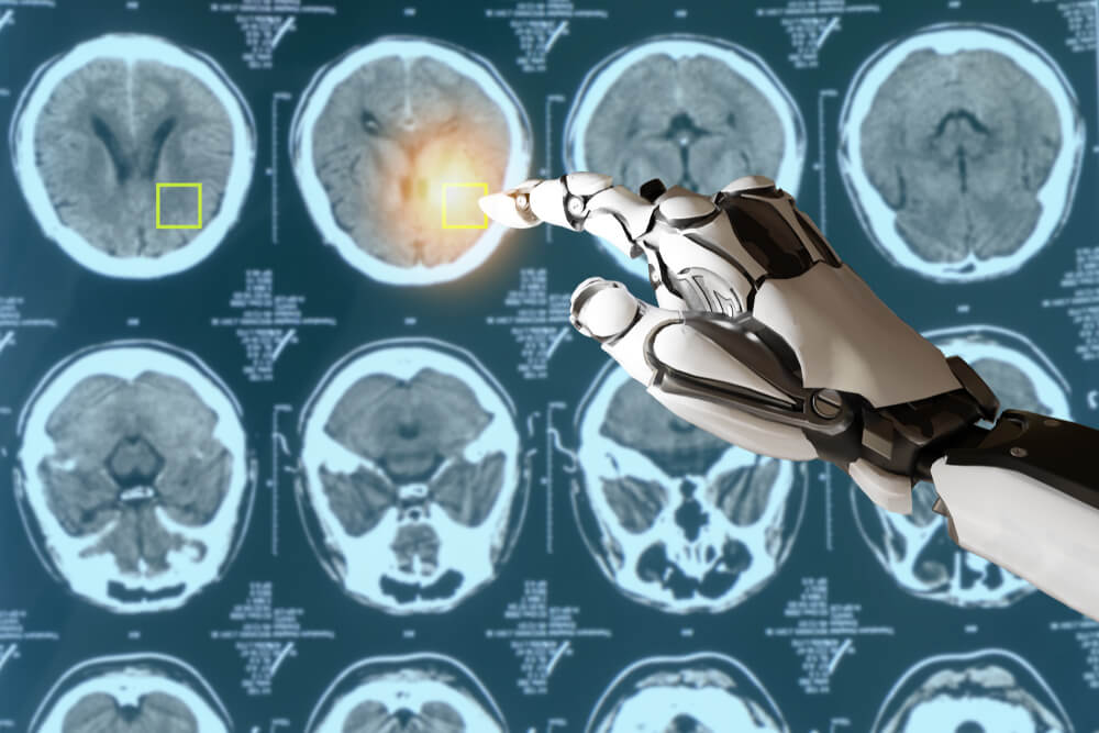
Medical imaging technology has transformed healthcare in recent decades. Various imaging modalities provide crucial visualization of the human body for accurate diagnosis, effective treatment planning, and precise intervention. From routine X-rays to complex PET scans, medical imaging enables health professionals to look inside the body non-invasively.
This blog post explores everything you need to know about the best medical imaging analysis. We will cover the key imaging modalities, their clinical applications, and technological advancements. Read on to gain valuable insights into this vital healthcare field.
X-Rays
X-ray imaging uses electromagnetic radiation to produce images of the body’s internal structures. X-rays pass through soft tissues but are absorbed by denser matter like bones. This contrast helps create detailed images.
X-rays are used to diagnose bone fractures, dental issues, pneumonia, tumors, and joint problems. They provide fast, painless evaluation at low costs. However, they do involve radiation exposure. Technological refinements now allow lower radiation doses.
Digital X-rays offer better image quality, faster processing, and easier data storage compared to traditional film X-rays. Advanced applications like tomosynthesis construct 3D views from multiple X-ray images. Overall, X-rays remain an indispensible first-line imaging tool.
Computed Tomography (CT) Scans
CT scans combine multiple X-ray images taken from different angles to generate cross-sectional views of the body. Powerful computer programs process the images.
CT scans provide more detailed organ visualization than plain X-rays. They are used to diagnose cancers, blood clots, head injuries, and cardiovascular diseases. CT angiography visualizes blood vessels, while CT colonography screens for colon cancer.
Newer CT scanners capture 128 or more simultaneous slices in sub-second timeframes. However, CT scans involve higher radiation exposure than X-rays. Proper dose optimization is important. CT remains integral for diverse diagnostic needs.
Magnetic Resonance Imaging (MRI)
MRI utilizes strong magnetic fields and radio waves to produce high-resolution, cross-sectional images of organs and tissues. It provides excellent soft tissue contrast.
MRI is advantageous for neurological, musculoskeletal, oncological, and cardiac imaging. It provides detailed evaluation without ionizing radiation. MRI has applications in tumor staging, degenerative spine disease, torn ligaments, stroke, and congenital heart defects.
Advanced 3.0T MRI systems allow faster imaging, better image quality, and thinner slices. Newer innovations like real-time MRI can track moving structures like the heart. Overall, MRI is invaluable for its versatility and non-invasive nature.
Ultrasound
Ultrasound uses soundwaves and their echoes to visualize internal anatomy. A transducer emits high-frequency soundwaves that reflect off tissue interfaces. These echoes are detected and processed into images.
Ultrasound provides real-time, dynamic imaging without radiation exposure. It is commonly used in obstetrics and gynecology to monitor pregnancy, evaluate the uterus and ovaries, and screen fetal development. Other applications include cardiac, liver, gallbladder, kidney, and vascular imaging.
Portable ultrasound devices allow imaging at bedside. Advanced techniques like 3D/4D ultrasound and contrast-enhanced ultrasound have expanded capabilities. Ultrasound is safe, inexpensive, and widely accessible.
Positron Emission Tomography (PET)
PET scanning involves injecting radiotracers into the body and using scanners to detect gamma rays emitted from tissues. It shows how well organs are functioning based on distribution of radiotracers.
PET is often combined with CT (PET-CT) to correlate anatomy and function. It is extensively used in oncology for cancer staging, detecting metastases, and evaluating treatment response. PET also has cardiology and neurology applications. However, PET is limited by high costs.
Newer scanners provide higher resolution with lower radiation doses. Innovations like PET-MRI integrate functional details from PET with excellent soft tissue contrast from MRI. PET continues to be an important molecular imaging modality.
Bone Densitometry
Bone densitometry measures bone mineral density (BMD) using specialized X-ray or ultrasound-based techniques like dual energy X-ray absorptiometry (DXA).
It is the established standard for diagnosing osteoporosis and assessing fracture risk. DXA scans are quick, painless and involve very low radiation. Quantitative data helps monitor changes over time and after treatment.
Vertebral fracture assessment (VFA) is performed using densitometers to detect vertebral compression fractures indicative of osteoporosis. Bone densitometry is vital for early osteoporosis diagnosis and management.
Nuclear Medicine Scans
Nuclear medicine uses radioactive tracers called radiopharmaceuticals that emit gamma rays. Special cameras detect signals from radiotracers concentrating in specific organs.
Examples include bone, thyroid, kidney, gallbladder, and stomach scans. Single photon emission computed tomography (SPECT) produces 3D images. Radiotracer mechanisms show organ function and pathology. However, radiation exposure needs monitoring.
Hybrid scans like SPECT-CT integrate anatomical and functional details. Advances continue, including new radiotracers for neuroimaging. Nuclear scans remain important for unique diagnostic information.
Fluoroscopy
Fluoroscopy uses continuous or pulsed X-rays to obtain real-time images of internal structures. The dynamic images guide interventional procedures.
Fluoroscopy enables visualization of catheters, stents, wires, and contrast agents during the procedure. Biopsies, joint injections, pacemaker implantation, and angiograms routinely use fluoroscopic guidance.
Newer digital systems allow image enhancement and lower radiation exposure. Fluoroscopy aids complex interventions but requires radiation precautions. Appropriate shielding and minimal exposure times are key.
The Future of Medical Imaging
Ongoing innovation aims to enhance imaging abilities, sensitivity, and efficiency while reducing costs and radiation exposure. Key trends include smaller and portable devices, advanced molecular imaging techniques, and imaging biomarkers.
Molecular imaging using techniques like PET visualizes cellular function. Imaging biomarkers can enable early disease detection and assess response to targeted therapies. Personalized precision diagnostics may become possible.
Other exciting areas include photoacoustic imaging, navigational procedures using augmented reality, and new tracers for neurodegenerative disorders. But regulations and access barriers need addressing first, along with clinician training in adopting new technologies.
Conclusion
Medical imaging technology has become indispensable in modern healthcare for reliable diagnosis, treatment planning, as well as interventional procedures. While X-rays and CT scans remain essential, newer modalities like MRI and PET add unique clinical value through their detailed functional assessments. Point-of-care ultrasound brings imaging directly to patients.
Ongoing progress aims to boost imaging capabilities while minimizing harms like radiation exposure. Artificial intelligence promises to aid image interpretation as well as quantification. But human expertise remains vital for sound clinical decision-making. Overall, diagnostic medical imaging usa is going through an exciting evolution that will continue enhancing patient care through precise visualization of diseases.


